The salamander kidney gives the final proof in 1938
 |
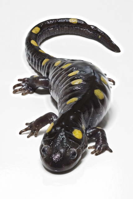
| A spotted salamander (Wikimedia Commons, see below) |
It was only in the 1930s that mainstream opinion accepted that the glomerulus was the problem in nephrotic syndrome – surprisingly late. But then it was only in 1924 that it was finally proven that the glomerulus is a filter, so perhaps we shouldn’t be surprised.
The origin of the confusion is easy to understand. On light microscopy the most consistent and prominent changes in nephrotic syndrome are the vacuoles in tubular cells. We now know this is a secondary response to their attempts to deal with excessive amounts of protein in the glomerular filtrate, but early pathologists assumed they were seeing the cause of the problem. Volhard and Fahr’s famous classification of Bright’s disease in 1914 made this assumption.
By 1940 better microscopy often showed some degree of change in the glomeruli, persuading many to the view that the glomerulus was the source. Bell pointed out histopathological evidence of abnormalities in the glomerulus in 1929, shortly after Wearn and Richard settled glomerular filtration, but arguments in the literature continued into the late 1930s (Waldherr and Ritz 1999). A glomerular origin of pathology was assumed by Ellis in 1942 for his ‘Type 2 nephritis’, although he does not acknowledge the definitive evidence from Germany more than two years previously.
Edmund Randerath and the salamander
 |
| Glomeruli in the salamander, drawn by Edmund Randerath (Copyright, see below) |
The salamander has two types of nephron, one of which opens into the coelomic cavity, the other being self-contained like mammalian nephrons. The pathologist Edmund Randerath, working in Dusseldorf, exploited this in a brilliantly simple experiment where he injected protein into the coelomic cavity of the salamander, and observed the effect on the two types of nephron. Only the tubules of those that communicated with the coelom showed vacuoles. The vacuoles then disappeared over a period of days as excess protein was cleared. The vacuoles were signs that they were actively dealing with the consequences of the excess protein, not the cause.
Waldherr and Ritz point out that Randerath was not widely credited or referenced despite decisively settling an important question. The war probably contributed to this, but it may also be that opinion had already accepted his explanation, so wasn’t surprised by his proof.
Further Reading
Volhard F, Fahr T. 1914. Die Bright’sche Nierenkrankheit, Klinik, Pathologie und Atlas. Springer, Berlin.
Fogazzi GB, Ritz E. 1998. Novel classification of glomerulonephritis in the monograph of Franz Volhard and Theodor Fahr. Nephrol Dial Transplant 13:2965-7
Waldherr R, Ritz E. 1999. Edmund Randerath (1899-1961): experimental proof for the glomerular origin of proteinuria. Kidney Int 56:1591-6.
Bell ET. 1929. Lipoid nephrosis. Am J Pathol 5:587-622. Excellent paper with case histories, good ilustrations; full paper free online.
The spectrum of glomerulonephritis (edrep.org)
Gross ML et al 2002. Intraperitoneal protein injection in the axolotl: the amphibian kidney as a novel model to study tubulointerstitial activation. Kidney Int 62:51-9. Gross et al picked up the salamander kidney idea over 75 years later, to test the ‘stressor’ effect of ingesting protein on tubular cells.
Image credits
Salamander – Camazine at en.wikipedia [CC BY-SA 3.0], via Wikimedia Commons
Slide by Edmund Randerath – from Waldherr R, Ritz E. 1999 with permission.