The shock of late, rapid dialysis – Glasgow, 1962
The early rotating drum dialysers had a large surface area, and because of the difficulties of doing each dialysis, coupled with the need for one-off vascular access each time until the Scribner shunt came into use, intervention was late, and dialysis sessions were infrequent and long. Patients often were not dialysed until they were very severely uraemic and obtunded, so perhaps it isn’t so surprising that only in 1961/62 were the late-dialysis or post-dialysis symptoms of dialysis disequilibrium noted.
 |
| The Hammersmith Hospital Usifroid dialysis machine, purchased to support its transplant programme in 1957. (Science Museum London, CC-BY-NC-SA licence as below) |
Arthur Kennedy was the doctor behind the introduction of dialysis in Glasgow in 1959. In 1962 he reported that some of their patients showed worsening or development of confusion, or headache. All recovered without mishap, but later reports described convulsions, coma and death. The underlying cause of renal failure and malignant hypertension may often have played a major part in these though.
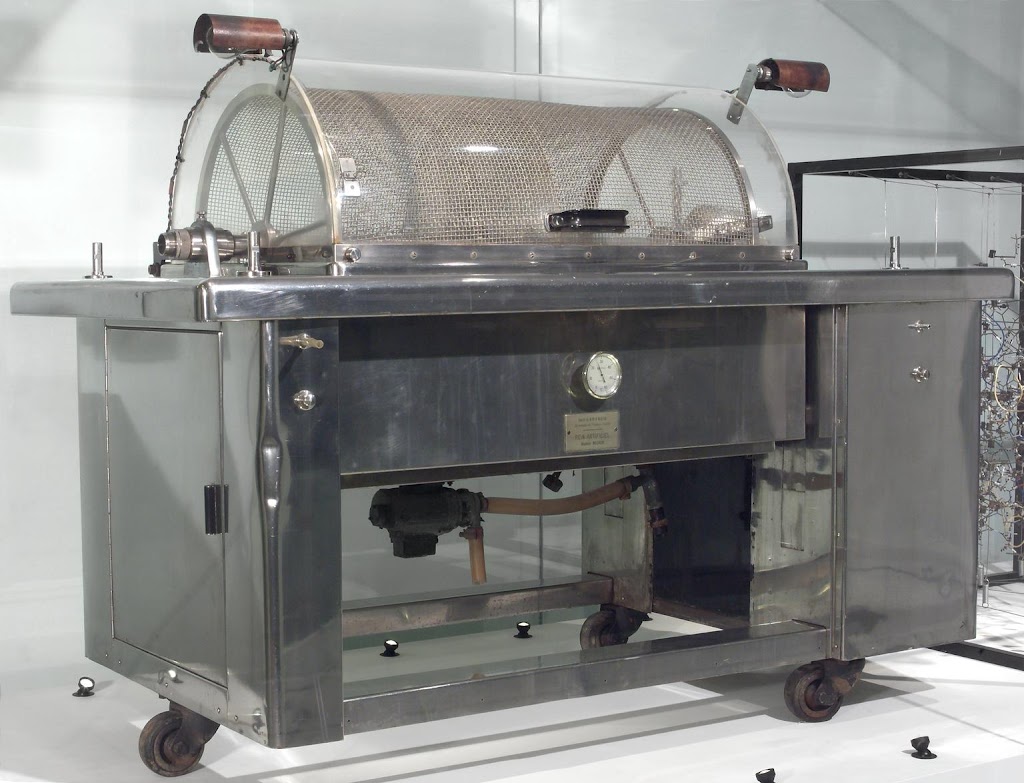
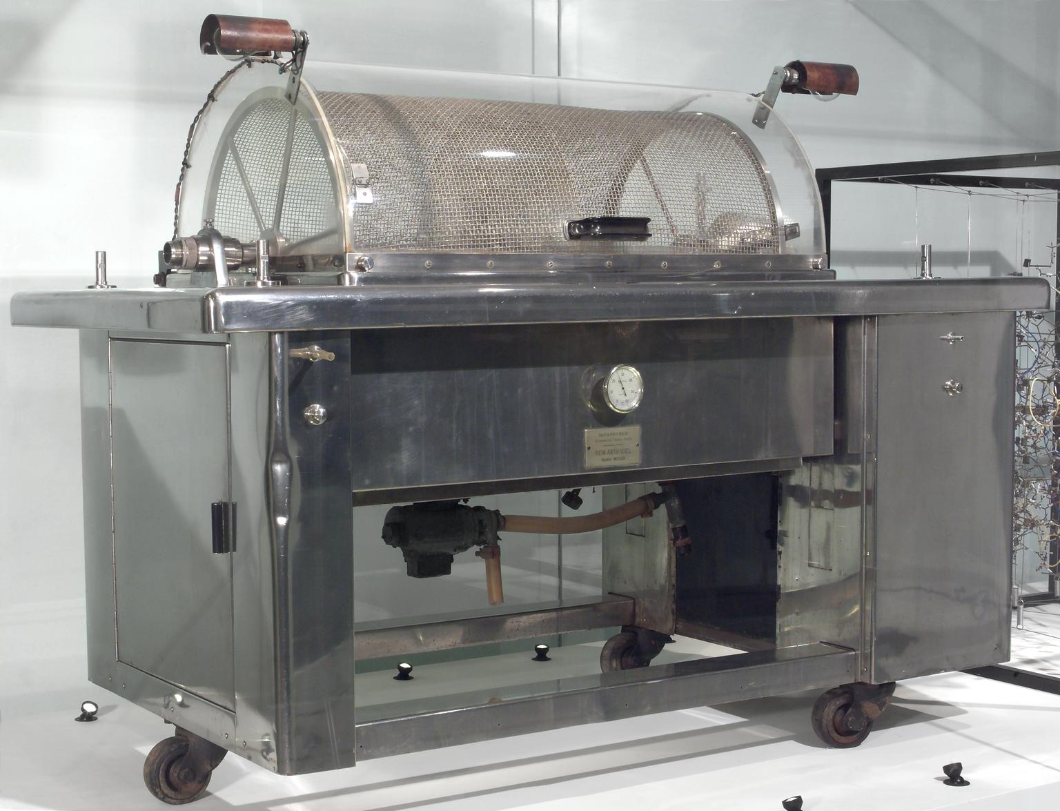
Kennedy’s machine was the Usifroid modification of Kolff’s rotating drum dialyser, first produced in 1955 for the Necker Hospital in Paris, in turn based on the Boston Kolff-Brigham machine. There were several similar machines in the UK; the Leeds Kolff-Brigham had been modified to give it a surface area of an astonishing 3.2 square metres. The one in Glasgow remained in use until 1979.
By reverse analogy with the use of intravenous urea in neurosurgery to reduce intracranial pressure, Kenedy hypothesized that lowering blood urea led to fluid accumulation in the brain.
Fig 1 shows the remarkable experiment of monitoring urea in cerebrospinal fluid by lumbar puncture before and after dialysis, and comparing it with blood values. These patients experienced huge changes in blood urea; from 80 to below 30 mmol/l in 4.5h in case 3 (495 to 190 mg/dl).
At the Royal Free in London, Kennedy’s observations were extended, and equilibration of urea found to be complete by 24h. Experiments in dogs (Sitprija and Holmes) confirmed that urea shifts drove a change in intracranial pressure.
 |
| Kennedy’s measurements of Urea in blood vs CSF (Lancet, as ref below, with permission) |
Symptoms of dialysis disequilibrium typically occur late in a long dialysis, or in the following hours. To avoid this we now introduce dialysis in short spells initially for very uraemic patients. Dialysis disequilibrium is more likely in patients with pre-existing brain disease or brain syndromes, in old or very young (=small) individuals, and in first treatments in very uraemic patients, where the osmolar shift is greatest.
In recent years, with more elderly patients or patients with pre-existing brain disease on dialysis, the lesser effects of even regular maintenance dialysis on mental function are being increasingly studied. The pathogenesis may be similar, more frequent dialysis (or peritoneal dialysis) often being recommended.
Further reading
Kennedy AC, AL Linton, JC Eaton. 1962. Urea levels in cerebrospinal fluid after haemodialysis. Lancet i:410-11
Zepeda-Orozco D, R Quigley. 2012. Dialysis disequilibrium syndrome. Pediatr Nephrol 27:2205-11 (open access)
Parsons F et al. 1961. Optimum time for dialysis in reversible renal failure. Lancet i 129-34. Describing modifications to increase surface area, and moves to intervene earlier in acute renal failure.
Rosen SM, K O’Connor, S Shaldon. 1964. Haemdialysis disequilibrium. Br Med J ii 672-5. A more extended study of CSF gradients of different molecules, and osmolality (open access)
Sitprija V, JH Holmes. 1962. Preliminary observations on the change in intracranial pressure and intraocular pressure during hemodialysis. JASAIO 8:300-8. In dogs, for intracranial pressure; but observing also that intraocular pressure rose in human patients (open access).
Arieff AI. 1994. Dialysis disequilibrium syndrome: current concepts on pathogenesis and prevention. Kidney Int 45:629-35 (open access)
Figure 1: Science Museum Group. Artificial kidney machine, France, 1955. Science Museum Group Collection Online. Accessed July 9, 2017. (Creative Commons licence).